Hi Dear Welcome to my blog. My blog is related to study which based upon Biology topics. Hope so my blog is helpful for you .
Hi Dear Welcome to my blog. My blog is related to study which based upon Biology topics. Hope so my blog is helpful for you .
Wednesday, 31 August 2022
Thursday, 18 August 2022
Explain Deformities of skeleton.
Deformities Of Skeleton
Human skeleton supports an upright body. Sometimes our skeleton system becomes weak and results in deformations. The causes of the deformations are available e.g.
Genetics Causes
Cleft Plate, a condition in which palatine processes of maxilla and palatine fail to fuse. The persistent opening between the oral and nasal cavity interferes with sucking. It can lead to inhalation of food into the lungs causing aspiration pneumonia.
Microcephaly, the small sized skull is caused by some genetic defect.
Arthritis covers over 100 different types of inflammatory of degenerative diseases that damage the joints .
Osteoarthritis, is the most common chronic arthritis, which is a degenerative joint disease also caused by genetic defect.
Hormonal Causes
Osteoporosis is a group of diseases in which bone resorption out paces bone deposit. In this case bone mass is reduced and chemical composition of the matrix remains normal. Osteoporosis mostly occurs in aged women, which is related to decreased estrogen level. Other factors which may contribute include, insufficient exercise, diet poor in calcium and protein, smoking. etc.
Estrogen replacement therapy offers the best protection against osteoporotic bone fractures.
Nutritional Causes
Osteomalcia (soft bone) includes a number of disorders in which the bones receive inadequate minerals. In this disease, calcium salts are not deposited and hence bones soften and weaken. Weight bearing bones of legs and pelvis bend and deform. The main symptom is the pain when height is put on affected bones.
Rickets is another disease in children with bowed legs and deformed pelvis. It is caused by deficiency of calcium in diet or vitamin 'D' deficiency. It is treated by vitamin 'D' fortified milk and exposing skin to sunlight to cure disorder.
Sunday, 14 August 2022
Write a note on joints.
Joints
Joints occurs where bones meet. They not only hold our skeleton together, but also gives it the mobility. Joints have three categories.
- Immovable joints
- Slightly movable joints
- Freely movable joints
- Hinge joints
- Ball and socket joint
- Fibrous Joints:
- Cartilaginous Joints:
- Synovial Joints:
- Hinge Joint:
- Ball & Socket joints:
Saturday, 13 August 2022
Explain commercial application of plant hormones.
Commercial Applications
Auxins
- Discovery of IAA led to the synthesis of wide range of compounds by chemists. The synthetic auxins are economical than IAA to produce and often more active because plants generally do not have necessary enzymes to break them down.
- NAA ( Naphthalene acetic acid)
- Indole propionic acid
- 2,4D (2,4 Dichloro phenoxy acetic acid)
- GA promote fruit setting e.g. in tangerines and pears and are used for growing seedless grapes. (Parthenocarpy) and also increase the berry size.
- GA3 is used in the brewing industry to stimulate a-amylase production in barely and this promotes malting.
- To delay ripening and improve storage life of bananas and grape fruits.
- Cytokinins delay aging of fresh leaf crops, such as cabbage and lettuce (delay of senescence) as well as keeping flowers fresh. They can also be used to break dormancy of some seeds.
- Abscisic acid can be sprayed on tree crops to regulate fruit drop at the end of the season. This removes the need for picking over a large time- span.
- Ethene induces flowering in pineapple. Stimulates ripening of tomatoes and citrus fruit. The commercial compound ethephon break down to release ethene in plants and is applied to rubber plant to stimulate the flow of latex.
Friday, 12 August 2022
Write down plants hormones.
Plant Hormones
Some of the special substances produced by the plants which influence the growth and plant responses to various stimuli are given below.
 |
| Plant Hormones |
(a) Auxins:
- In stem, promote cell enlargement in region behind apex. Promote cell division in cambium.
- In roots, promote growth at very low concentration. Inhabit growth at higher concentration e.g. geotropism. Promote growth of roots from cuttings and calluses.
- Promote bud initiation in shoots but sometimes antagonistic to cytokinins and is inhibitory.
- Cause delay in leaf senescence in a few species.
- Promote cell enlargement in the presence of auxins. Also promote cell division in apical meristem and cambium.
- Promote "bolting" of some rosette plants.
- Promote bud initiation in shoots of chrysanthemum callus.
- Cause delay in leaf senescence in few species.
- Break bud and seed dormancy.
- Promote leaf growth and fruit growth.
- Promote stem growth by cell division in apical meristem and cambium.
- Promote stomatal opening.
- Cause delay in leaf senescence.
- Inhibitory primary root growth.
- Promote lateral root growth.
- Promote bud initiation and leaf growth.
- Promote bud and seed dormancy.
- Promote flowering in short day plants, and inhibits in long day plants.
- Sometimes promotes leaf senescence.
- Promote abscission.
- Promote closing of stomata under condition of water stress.
- Inhibits root growth.
- Break dormancy of bud.
- Promotes fruit ripening.
- Promotes flowering in pineapple.
- Inhibits stem growth.
Monday, 8 August 2022
Write a note on Ear.
Ear
Hearing is important as vision. Our ear helps us in hearing and also to maintain the balance or equilibrium of our body.
- External Ear
- Middle Ear
- Inner Ear
- Vestibule
- Semicircular canals
- Cochlea
Sunday, 7 August 2022
Write a note on Spinal Cord?
Spinal Cord
The spinal cord is in fact a tubular bundles of nerves. It starts from brain stem and extends to lower back. Like, brain, spinal cord is also covered by meninges. The vertebral column surrounds and protect spinal cord.
The outer region of spinal cord is made of white matter (containing myelinated axons). The central region is butterfly shaped that surrounds the central canal. Its made of grey matter (containing neuron cell bodies).
31 pairs of spinal nerves arise along spinal cord. These are " Mixed Nerves" because each contains axons of both sensory and motor neurons.
At the point where a spinal nerve arises from spinal cord, There are two roots of spinal nerve.
- The dorsal root contains sensory axons and ganglion where cell bodies are located.
- The ventral root contains axons of motor neurons.
- It serves as a link between body parts and brain. Spinal cord transmits nerve impulses from body parts to brain and from brain to body parts.
- Spinal cord also acts as coordinator, responsible for some simple reflexes.
Who is the largest part of brain & write its part?
Brain
Forebrain is the largest parts of brain. It is most highly developed in humans.
FOREBRAIN
Parts:
There three parts of forebrain.
- Thalamus
- Hypothalamus
- Cerebrum
Lies just below the cerebrum. It serves as a relay center between various parts of brain and spinal cord. It also receives and modifies sensory impulses (except nose) before they travel to cerebrum. Thalamus is also involved in pain perception and consciousness (sleep and awakening).
Hypothalamus:
Lies above midbrain and just below thalamus. In humans, it is roughly the size of an almond. One of the most important functions of hypothalamus is to link nervous system and endocrine system. It control the secretions of pituitary gland. It also controls feelings such as rage, pain, pleasure and sorrow.
Cerebrum:
It is the largest parts of forebrain. It controls skeletal muscles, thinking, intelligence and emotions. It is divided into two cerebral hemisphere.
Cerebral hemisphere:
The anterior parts of cerebral hemisphere are called Olfactory bulbs which receive impulses from olfactory nerves and create the sensation of smell.
Upper layer:
The upper layer of cerebral hemispheres i.e. cerebral cortex consists of grey matter. The grey matter of nervous system consists of cell bodies and non myelinated axons.
Lower layer:
This layer consists of white matter. The white matter of nervous system consists of myelinated axons.
Cerebral cortex has large surface area and is folded in order to fit in skull. It is divided into four lobes.
Explain Neuron & Write three classification of nerve.
Neuron
Nerve cell or neuron is the unit of the nervous system. The human nervous system consists of billions of neurons plus supporting cell. Neuron are the specialized cells that are able to conduct nerve impulses from receptors to coordinators and from coordinators to effectors. In this way the communicate with each other and with other types of body cell.
Location:
The nucleus and most of the cytoplasm of neurons is located in the cell body.
Dendrites & Axons:
Different processes extend out from the cell body. These are called dendrites and axons.
Dendrites conduct impulses towards cell body and axons conduct impulses away from the cell body.
Schwann cells:
Schwann cells are special neurological cells located at regular intervals along axons, In some neurons Schwann cell secrete fatty layer called myelin sheath.
Nodes of Ranvier:
Between the areas of myelin on an axon, there are no myelinated points called Nodes of Ranvier.
Saltatory:
Myelin sheath is an insulator so the membrane coated with this sheath does not conduct nerve impulse. In such impulses are called saltatory (jumping) impulses. This increases the speed of nerve impulse.
On the basis of the function, neuron are of three types.
- Sensory neurons
- Interneurons
- Motor neurons
Conduct sensory information (nerve impulse) from receptors towards the CNS. Sensory neurons have on dendrite and one axon.
Interneuron:
From brain and spinal cord. They receive information, interpret them and stimulate motor neurons. They have many dendrites and axons.
Motoneuron:
Carry information from interneurons to muscle or glands (effectors). They have many dendrites but only one axon.
Nerve
A nerve means the union of several axons that are enveloped by covering made of lipid.
Classification:
- Sensory nerve
- Motor nerve
- Mixed nerve
Contain the axons of sensory neurons only.
Motor nerve:
Contain the axons of motor neurons only.
Mixed neuron:
Contain the axons of both i.e. sensory and motor neurons.
Saturday, 6 August 2022
Write down the steps of functionality of kidney.
Functioning of kidney
The main function of kidney is urine formation, which takes place in three steps.
- Pressure Filtration
- Selective Reabsorption
- Tubular Secretion
Pressure Filtration:
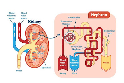
When blood enters the kidney via the renal artery, it goes to many arterioles, and then to the glomerulus . The pressure of blood is very high and so most of the water, salt, glucose and urea of blood is forced out of glomerulus capillaries. This material passes into the Bowman's capsule and is now called Glomerulus Filtrate.
Selective Reabsorption:
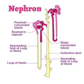
The second step is the selective reabsorption. In this step about 99% of the glomerulus filtrate is reabsorbed into the blood capillaries surrounding renal tubule. It occurs through osmosis, diffusion and active transport. Some water and most of the glucose is reabsorbed from the proximal convoluted tubule. Here, salts are reabsorbed by active transport and then follows by osmosis. The descending limb of loop of Henle allows the reabsorption of water while the ascending limb of loop of Henle allows the reabsorption of salts. The distal convoluted tubule again allows the reabsorption of water into the blood.
Tubular Secretion:
The third step is tubular secretion . Different ions, creatinine, urea etc. are secreted from blood into the filtrate in renal tubule. This is done to maintain blood at normal pH (7.35 to7.45).
Define urine & Explain osmoregulatory function of kidney.
Urine
The filtrate present in renal tubules is known as urine. It moves into collecting ducts and then into pelvis.
Osmoregulatory function of kidney
Osmoregulation:
Is defined as the regulation of the concentration of the water and salt in blood and other body fluids. Kidney plays important role in osmoregulation by regulating the water fluids whereas excess intake of water dilutes them.
Hypotonic:
When there is excess water in body fluids, kidney from dilutes (Hypotonic) urine. For this purpose, kidney filter more water from glomerulus capillaries into Bowman's capsule. Similarly less water is reabsorbed and abundant dilute urine is produced. It brings down the volume of the body fluids to normal.
Hypertonic:
When there is shortage of water in body fluids, kidney filter less water from glomerulus capillaries and the rate of reabsorption of water is increased. Less filtration and more reabsorption of water produce small amount of concentration (hypertonic) urine. It increases the volume of body fluids to normal. This whole process is under hormonal control.
Previous work
Hypotonic & Hypertonic
Why Kidney Stone happen & what is Renal Failure?
Kidney Stone
When urine becomes concentrated, crystals of many salts eg calcium oxalate, calcium and ammonium phosphate, uric acid etc. are formed in it. Such large crystals cannot pass in urine and form hard deposits called Kidney Stone.
Most stones start in kidney. Some may travel to ureter or urinary bladder.
Causes:
The major causes of kidney stones are age, diet (containing more green vegetables, salts, vitamins C,D), recurring urinary tract infection , less intake of water, and alcohol consumption.
Symptoms:
The symptoms of kidney stones include severe pain in kidney or in lower abdomen, vomiting, frequent urination and foul-smelling urine with blood and pus.
About 90% of all kidney stones can pass through the urinary system by drinking plenty of water. In surgical treatment, the affected area is open and stones are removed.
Lithotripsy:
Lithotripsy is another method of removal of kidney stones. In this methods, non electrical shock waves from outside are bombarded on the stone in the urinary system. Waves hit the dense stones and break them. Stones become sand-like and are passed through urine.
Kidney(Renal) Failure
Kidney failure means a complete or partial failure of kidneys to function.
Diabetes mellitus and hypertension are the leading causes of kidney failure. In certain cases, sudden interruption in the blood supply to kidney and drug overdoses may also result in kidney failure.
Symptoms:
The symptoms of kidney failure is the high level of urea and other wastes in blood, which can result in vomiting, nausea, weight loss, frequent urination and blood in urine. Excess fluids in body may also cause swelling in legs, feet face and shortness of breath.
Treatment:
The kidney failure is treated with dialysis and kidney transplant.
Friday, 5 August 2022
Write a note on Dialysis.
Dialysis
Dialysis means cleaning of blood by artificial ways.
Methods:
There are two methods of dialysis.
- Peritoneal Dialysis
- Haemo Dialysis
In this type of dialysis, the dialysis fluid is pumped for a time into the peritoneal cavity which is the space around gut. This cavity is lined by peritoneum. Peritoneum contains blood vessels. When we place dialysis fluid in peritoneal cavity, waste materials from peritoneal blood vessels diffuse into the dialysis fluid, which is then drained out. This type of dialysis can be performed at home, but must be done every day.
Haemodialysis:
In haemodialysis, patient's blood is pumped through an apparatus called Dialyzer. The dialyzer contains long tubes, the walls of which act as semi-permeable membranes. Blood flows through the tubes while dialysis fluid flows around the tubes. Extra water and wastes moves from blood into the dialysis fluid. The cleansed blood is then returned back to body. The haemodialysis treatments are typically given in the dialysis center.
Write a note on structure and function of kidney.
Structure of Kidney
Colour of kidney:
Kidney are dark red bean shaped organ.
Size of kidney:
Each kidney is 10cm long, 5cm width thick and weights about 120 grams.
Location:
They are placed against the back wall of abdominal cavity just below diaphragm , one on either side of the vertebral column. They are protected by the last 2 ribs. The left kidney is little higher than the right.
Hilus:
The concave side of the kidney faces the vertebral column. There is a depression called Hilus, near the Centre of the concave area of kidney.
Kidney shows two regions
Renal cortex:
is the outer part of the kidney and it is dark red in colour.
Renal medulla:
is the inner part of the kidney and is pale red in colour. Renal medulla consists of several cone shaped areas called renal Pyramids. Renal pyramids projects into a funnel-shaped cavity called renal Pelvis which is the base of ureter.
 |
| Structure of kidney |
Osmotic adjustments in plants & Explain its three groups.
Osmotic adjustment in plants
On the basis of available amount of water and salt, plants divided into three groups.
Hydrophytes:
Those plants which live partially submerge or completely in freshwater. Such plants do not face the problem of water shortage. They have developed mechanisms for the removal of extra water from their cells. Hydrophytes have broad leaves with a large number of stomata on their upper surfaces. This characteristics helps them to remove the extra amount of water.
Example:
Water lily.
Xerophytes:
They live in dry environments. They possess thick, waxy cuticle over their epidermis to reduce water loss from internal tissues. They have less number of stomata to reduce the rate of transpiration. Such plants have deep roots to absorb maximum water from soil.
Example:
Cacti (singular cactus).
Halophytes:
They live in sea water and are adapted to salty environment. Salts enter in the bodies of such plants due to their higher concentration in sea water.
Example:
Sea grass.

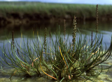
Halophytes plants
Previous work
What is Genetic Engineering? Explain.
Genetic engineering , also known as genetic modification or genetic manipulation, is a set of techniques that involve the direct manipulat...
-
Osmotic adjustment in plants On the basis of available amount of water and salt, plants divided into three groups. Hydrophytes: ...
-
Neuron Nerve cell or neuron is the unit of the nervous system. The human nervous system consists of billions of neurons plus supporting cel...
-
Lungs All the alveoli on one side constitute a lung. There is a pair of lungs in the thoracic cavity. The chest wall is made up of 12 pairs...


.png)


.png)