Saturday 6 August 2022

Define urine & Explain osmoregulatory function of kidney.

 Urine

The filtrate present in renal tubules is known as urine. It moves into collecting ducts and then into pelvis.

Osmoregulatory function of kidney

Osmoregulation:

                                  Is defined as the regulation of the concentration of the water and salt in blood and other body fluids. Kidney plays important role in osmoregulation by regulating the water fluids whereas excess intake of water dilutes them.

Osmoregulation of kidney


Hypotonic:

                       When there is excess water in body fluids, kidney from dilutes (Hypotonic) urine. For this purpose, kidney filter more water from glomerulus capillaries into Bowman's capsule. Similarly less water is reabsorbed and abundant dilute urine is produced. It brings down the volume of the body fluids to normal.

Hypertonic:

                        When there is shortage of water in body fluids, kidney filter less water from glomerulus capillaries and the rate of reabsorption of water is increased. Less filtration and more reabsorption of water produce small amount of concentration (hypertonic) urine. It increases the volume of body fluids to normal. This whole process is under hormonal control.

Hypotonic & Hypertonic


Previous work

Why Kidney Stone happen & what is Renal Failure?

 Kidney Stone

When urine becomes concentrated, crystals of many salts eg calcium oxalate, calcium and ammonium phosphate, uric acid etc. are formed in it. Such large crystals cannot pass in urine and form hard deposits called Kidney Stone.

kidney stones


Most stones start in kidney. Some may travel to ureter or urinary bladder.

Causes:

               The major causes of kidney stones are age, diet (containing more green vegetables, salts, vitamins C,D), recurring urinary tract infection , less intake of water, and alcohol consumption.

Symptoms:

                      The symptoms of kidney stones include severe pain in kidney or in lower abdomen, vomiting, frequent urination and foul-smelling urine with blood and pus. 

About 90% of all kidney stones can pass through the urinary system by drinking plenty of water. In surgical treatment, the affected area is open and stones are removed.

Lithotripsy:

                       Lithotripsy is another method of removal of kidney stones. In this methods, non electrical shock waves from outside are bombarded  on the stone in the urinary system. Waves hit the dense stones and break them. Stones become sand-like and are passed through urine.

Kidney(Renal) Failure

Kidney failure means a complete or partial failure of kidneys to function.

Renal Failure
Causes:

               Diabetes mellitus and hypertension are the leading causes of kidney failure. In certain cases, sudden interruption in the blood supply to kidney and drug overdoses may also result in kidney failure.

Symptoms:

                       The symptoms of kidney failure is the high level of urea and other wastes in blood, which can result in vomiting, nausea, weight loss, frequent urination and blood in urine. Excess fluids in body may also cause swelling in legs, feet face and shortness of breath.

Treatment:

                       The kidney failure is treated with dialysis and kidney transplant.


Previous work

Friday 5 August 2022

Write a note on Dialysis.

 Dialysis

Dialysis means cleaning of blood by artificial ways.

Methods:

                There are two methods of dialysis.

  1. Peritoneal Dialysis
  2. Haemo Dialysis
Peritoneal Dialysis:

                                        In this type of dialysis, the dialysis fluid is pumped for a time into the peritoneal cavity which is the space around gut. This cavity is lined by peritoneum. Peritoneum contains blood vessels. When we place dialysis fluid in peritoneal cavity, waste materials from peritoneal blood vessels diffuse into the dialysis fluid, which is then drained out. This type of dialysis can be performed at home, but must be done every day.


Haemodialysis:

                                In haemodialysis, patient's blood is pumped through an apparatus called Dialyzer. The dialyzer contains long tubes, the walls of which act as semi-permeable membranes. Blood flows through the tubes while dialysis fluid flows around the tubes. Extra water and wastes moves from blood into the dialysis fluid. The cleansed blood is then returned back to body. The haemodialysis treatments are typically given in the dialysis center.


Previous work

Write a note on structure and function of kidney.

 Structure of Kidney

Colour of kidney:

                                   Kidney are dark red bean shaped organ.

Size of kidney: 

                             Each kidney is 10cm long, 5cm width thick and weights about 120 grams.

Location:

                   They are placed against the back wall of abdominal cavity just below diaphragm , one on either side of the vertebral column. They are protected by the last 2 ribs. The left kidney is little higher than the right.

Hilus: 

            The concave side of the kidney faces the vertebral column. There is a depression called Hilus, near the Centre of  the concave area of kidney. 

Kidney shows two regions

Renal cortex:

                          is the outer part of the kidney and it is dark red in colour.

Renal medulla:

                                is the inner part of the kidney and is pale red in colour. Renal medulla consists of several cone shaped areas called renal Pyramids. Renal pyramids projects into a funnel-shaped cavity called renal Pelvis which is the base of ureter.

Structure of kidney


Function of Kidney

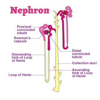
The function unit of kidney is called nephron. There are over one millions nephrons in each kidney. 
Parts of Nephron:
                                    There are two parts of nephron i.e. Renal Corpuscle and Renal Tubule.

Renal corpuscle is not tubular and has two parts i.e. glomerulus and Bowman's capsule. Glomerulus is a network of capillaries while Bowman's capsule is a cap shaped structure that encloses glomerulus.

Renal Tubule:
                            It is the part of nephron which starts after Bowman's capsule. Its first portion is called the proximal convoluted tubule. Next portion is U-shaped and is called Loop of Henle. The last portion of renal tubule is called the Distal convoluted tubule. 
Collecting Duct:
                                 The distal convoluted tubule of many nephrons open in a single collecting duct. Many collecting ducts join together to form several hundred Papillary Ducts which drain into renal pelvis.

Osmotic adjustments in plants & Explain its three groups.


Osmotic adjustment in plants

On the basis of  available amount of water and salt, plants divided into three groups.

Hydrophytes:

                           Those plants which live partially submerge or completely in freshwater. Such plants do not face the problem of water shortage. They have developed mechanisms for the removal of extra water from their cells. Hydrophytes have broad leaves with a large number of stomata on their upper surfaces. This characteristics helps them to remove the extra amount of water.

Example:

                  Water lily.

Hydrophytes plants


Xerophytes: 

                        They live in dry environments. They possess thick, waxy cuticle over their epidermis to reduce water loss from internal tissues. They have less number of stomata to reduce the rate of transpiration. Such plants have deep roots to absorb maximum water from soil. 

Example:

                  Cacti (singular cactus).

Xerophytes plants


Halophytes:

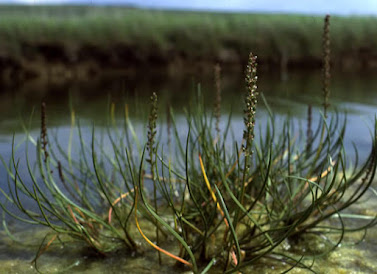
                         They live in sea water and are adapted to salty environment. Salts enter in the bodies of such plants due to their higher concentration in sea water. 

Example:

                  Sea grass.

Halophytes plants


Previous work

Saturday 30 July 2022

Why left lung is small and right lung is large ? Explain.

 Lungs


All the alveoli on one side constitute a lung. There is a pair of lungs in the thoracic cavity. The chest wall is made up of 12 pairs of ribs and the rib muscles called intercoastal muscles.


Diaphragm:

                        A thick muscular structure, is called diaphragm, is present below the lungs.

Left & Right lungs

The left lung is slightly smaller and has two lobes  and right lung is bigger and has three lobes. They are spongy and elastic organs. The lungs also have blood vessels that are the branches of the pulmonary arteries and veins. Each lung is enclosed by two membranes called outer pleural membrane and inner pleural membrane. 

The membranes enclose a fluid which provides lubrication for the free expanding and contracting of lungs.

What is air passageway?

 Air Passageway

The air passageway consists of the parts through which the outside air comes in the lungs and after the exchange of gases it goes out. This passage of air  consists of the following parts.


The nose encloses the Nasal Cavity.

Nostrils:

                 It opens  to the outside through the openings called the Nostrils.


Division of Nasal Cavity:

                                                  The nasal cavity is divided into two portions by a wall. Each portion is lined by fine hairs and mucous which filter the dust particles from the air. The mucous also moistens and warm the incoming air and keeps its temperature nearly equal to that of the body.

Internal Nostrils: 

                                   The nasal cavity opens into the pharynx by means of two small openings called internal nostrils.  

Pharynx: 

                  Pharynx is the muscular passage and is common to both food and air. It extends to the opening of oesophagus and the larynx. The air goes from the pharynx into the larynx.

Glottis: 

               It is a narrow opening at the floor of pharynx which leads into larynx.


Larynx: 

                The larynx is a box, made of cartilage. It is present between pharynx and trachea. It is also called Voice Box. 

Vocal cords: 

                        Two pairs of fibrous brands called vocal cords are stretched across the larynx. The vocal cords vibrate when the air passes through them. This vibration produces sounds. 

Wind pipe:

                     Larynx continues to the trachea, which is also called the wind pipe. It is about 12cm long tube which lies in the front of the oesophagus. There are C-shaped cartilagenous rings in the wall of Trachea. The cartilages keeps the trachea from collapsing even when there is no air in it.

Bronchi:
                       On entering the chest cavity , the trachea divides into smaller tubes called Bronchi (singular bronchus).
Bronchioles:
                         The bronchi continue dividing in the lungs until they make several fine tubes called Bronchioles.
Alveolar ducts: 
                                                            The bronchioles end as fine tubules called alveolar ducts.
Alveoli: 
             Each alveolar duct opens into a cluster of pouches called Alveoli.

What is Genetic Engineering? Explain.

  Genetic engineering , also known as genetic modification or genetic manipulation, is a set of techniques that involve the direct manipulat...